


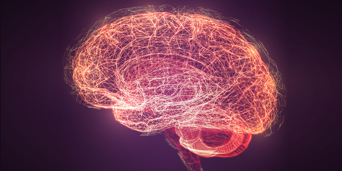
A team of scientists recently aimed to better understand consciousness and its pathologies by studying the neural activity of patients with disorders of consciousness and healthy volunteers using brain imaging technology. They identified two crucial brain circuits implicated in consciousness. The results of the study have been published in Human Brain Mapping.
Consciousness is a complex and subjective experience, and there is still much debate among scientists and philosophers about its nature and origin. However, in clinical settings, doctors treating patients with severe brain injuries and disorders of consciousness need to find ways to help their patients, regardless of the exact definition of consciousness. The authors of the new study sought to better understand the mechanisms behind the pathological loss of consciousness and its recovery, as well as to have reliable ways to assess the state of the patients.
“In recent years, many studies have tried to objectively assess levels of consciousness using various neuroimaging techniques. While these studies have improved how we diagnose patients with disorders of consciousness, they haven’t fully explained how consciousness comes about,” explained study author Jitka Annen, a postdoctoral researcher at the Coma Science Group at the University of Liege.
“With this work, we wanted to characterize how a stimulus (or perturbation in the ongoing activity) affects the flow of neural activity in different states of reduced consciousness after brain injury. By doing so, we gain insights in how regions receive activity from and broadcast activity to other brain regions, or how some subjects can sustain consciousness while in others cannot. In brief, we attempted to better understand how information flow differs in various states of consciousness.”
To achieve these goals, the researchers used neuroimaging methods, particularly functional magnetic resonance imaging (fMRI), to study brain activity in patients with disorders of consciousness and healthy volunteers. They focused on how external perturbations, such as sensory stimuli or artificial stimulation, propagate through the brain in different states of reduced consciousness. By observing these brain dynamics, they aimed to understand how information flows in the brain and how different regions interact with each other during consciousness and altered states of consciousness.
For the study, they included subjects with disorders of consciousness after severe brain injury (such as those in a minimally conscious state and unresponsive wakefulness syndrome) and healthy control volunteers without neurological problems. They performed behavioral assessments and used glucose PET scans to confirm the diagnosis of disorders of consciousness patients. The researchers acquired structural and functional MRI data from all participants.
Using the fMRI data, the team studied the propagation of endogenous (spontaneous) and in-silico exogenous (model-based) perturbations in the brains of patients with disorders of consciousness. They estimated directed and causal interactions between brain regions based on the resting-state fMRI data and fitted them to a linear model of activity propagation.
The researchers found that patients with disorders of consciousness had malfunctioning neural circuits in their brains. These circuits failed to convey and integrate information properly, leading to a lack of consciousness.
In healthy individuals, they found that specific brain regions were responsible for broadcasting and receiving information, which is crucial for conscious perception. However, in patients with unresponsive wakefulness syndrome (UWS), the brain’s ability to transmit and integrate information was severely impaired. They observed a rapid decay of brain activity and reduced connectivity between brain regions, leading to a lack of conscious perception.
For patients in a minimally conscious state, who had partially recovered consciousness, some of these brain functions showed improvement, indicating that certain brain regions were regaining their ability to process and integrate information.
“The brains of patients with a disorder of consciousness have hampered capacity to respond to events,” Annen told PsyPost. “And when events reach the cortex, their activity does not propagate successfully to other cortical areas for further processing. In healthy volunteers, brain regions are embedded in specialized networks.”
“Specifically, the posterior part of brain (back of brain, receiving sensory information) and thalamus (an inner part of brain functioning as gateway between the body and the brain) are specialized to integrate the flow of activity while frontal and temporal (lateral) cortical regions (responsible for various cognitive processes) are specialized to propagate activity of events.”
“This functional specialization seems to be lost in unconscious patients. All regions behave similarly, and activity is not sustained long enough to be processed by the relevant regions. In patients showing minimal consciousness, these properties of functional neural processing recover.”
The study also found that the brain’s glucose consumption (as measured via PET scans) was directly related to how well it processed stimuli. In other words, when the brain’s glucose metabolism was higher, it showed a better ability to respond to and process information effectively. This suggests that the brain’s ability to process information is closely linked to its overall health.
“Stimulus processing was proportional to the sugar consumption of the brain, a very robust measure of neural integrity,” Annen said. “While this could be considered to be expected, I find it remarkable that the relation is so direct especially given that the glucose consumption data comes from a totally different type of brain scan in the same subjects.”
The study provides important insights into the mechanisms behind consciousness and how they can be affected by brain injuries. Understanding these processes could potentially help clinicians find better ways to treat and support patients with disorders of consciousness. But as with all research, the study includes some caveats.
“This work was performed in the framework of the Human Brain Project and would not have been possible without Dr. Rajanikant Panda, and our collaborators at the University Pompeu Fabra Barcelona (Spain), Ane López-González and Gorka Zamora-López,” she added.
The study, “Whole-brain analyses indicate the impairment of posterior integration and thalamo-frontotemporal broadcasting in disorders of consciousness“, was authored by Rajanikant Panda, Ane López-González, Matthieu Gilson, Olivia Gosseries, Aurore Thibaut, Gianluca Frasso, Benedetta Cecconi, Anira Escrichs, Coma Science Group Collaborators, Gustavo Deco, Steven Laureys, Gorka Zamora-López, and Jitka Annen.









Discussion about this post